Understanding Amyloidosis
Amyloidosis is a rare group of diseases characterized by the abnormal buildup of proteins called amyloids in various tissues and organs throughout the body. This can lead to organ dysfunction and potentially life-threatening complications if left untreated.
What is Fat Pad Biopsy?
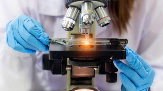
A fat pad biopsy is a minimally invasive procedure used to obtain a small sample of fat tissue from just beneath the skin. This tissue sample can then be examined under a microscope to detect the presence of amyloid deposits, aiding in the diagnosis of amyloidosis.
Indications for Fat Pad Biopsy
Fat pad biopsy may be recommended in patients with suspected amyloidosis based on clinical symptoms, laboratory tests, or imaging findings. It is particularly useful when other diagnostic methods, such as tissue biopsy of affected organs, are not feasible or inconclusive.
How is Fat Pad Biopsy Performed?
During a fat pad biopsy, a small incision is made in the skin, usually on the abdomen or near the knee. A specialized needle is then inserted into the subcutaneous fat layer to obtain a tissue sample. The procedure is typically performed under local anesthesia and takes only a few minutes to complete.
Risks and Complications
Fat pad biopsy is considered a safe procedure with minimal risks. However, as with any invasive procedure, there is a small risk of bleeding, infection, or damage to surrounding tissues. These risks are generally low and can be minimized by following proper surgical techniques and post-procedure care.
Interpreting Fat Pad Biopsy Results
The tissue sample obtained from a fat pad biopsy is examined under a microscope by a pathologist to look for characteristic features of amyloid deposits, such as abnormal protein accumulation between fat cells. A positive result indicates the presence of amyloidosis, while a negative result does not necessarily rule out the disease.
Types of Amyloidosis Detected by Fat Pad Biopsy
Fat pad biopsy can detect various types of amyloidosis, including AL amyloidosis (caused by abnormal production of immunoglobulin light chains), AA amyloidosis (associated with chronic inflammatory conditions), and ATTR amyloidosis (due to abnormal transthyretin protein).
Limitations of Fat Pad Biopsy
While fat pad biopsy is a valuable diagnostic tool for amyloidosis, it may not always provide definitive results. In some cases, amyloid deposits may be sparse or difficult to detect in the tissue sample obtained, leading to false-negative results. Additionally, fat pad biopsy cannot differentiate between different types of amyloidosis, requiring further testing for subtype classification.
Clinical Utility of Fat Pad Biopsy
Despite its limitations, fat pad biopsy remains an important tool in the diagnosis and management of amyloidosis. It is relatively safe, minimally invasive, and can provide valuable diagnostic information, especially in cases where other diagnostic methods are inconclusive or unavailable.
Combining Fat Pad Biopsy with Other Diagnostic Tests
In many cases, fat pad biopsy is used in conjunction with other diagnostic tests, such as serum protein electrophoresis, imaging studies, and tissue biopsies of affected organs, to confirm the diagnosis of amyloidosis and determine its underlying cause.
Treatment Implications
Once amyloidosis is diagnosed, treatment strategies can be tailored to the specific type and severity of the disease. These may include chemotherapy, immunomodulatory drugs, organ transplantation, or supportive care measures to manage symptoms and improve quality of life.
Prognosis and Follow-up
The prognosis for patients with amyloidosis depends on various factors, including the type and extent of organ involvement, response to treatment, and presence of comorbidities. Regular follow-up visits with a healthcare provider are essential for monitoring disease progression, adjusting treatment as needed, and addressing any complications that may arise.
Conclusion
Fat pad biopsy is a valuable diagnostic tool in the evaluation of patients with suspected amyloidosis. It offers a relatively simple, safe, and minimally invasive means of detecting amyloid deposits in subcutaneous fat tissue, helping to confirm the diagnosis and guide further management decisions. While fat pad biopsy has its limitations, when used in combination with other diagnostic tests, it can provide valuable insights into the underlying cause and severity of amyloidosis, ultimately improving patient outcomes.


